The Killian-Jamieson diverticulum was first identified in 1983 by Ekberg and colleagues. It is located in the anterior wall of the cervical esophagus, distal to the cricopharyngeal muscle, and it can be diagnosed by endoscopy. Uncertain pathogenesis characterizes this unusual esophageal diverticulum.
In the cervical esophagus, Killian Jamieson’s Diverticulum arises laterally from the longitudinal muscle and inferiorly from the cricopharyngeal musculature. It is different from the more common Zenker’s diverticulum, which forms in the middle of the back wall of the esophagus, just above the cricopharyngeal muscle (this area is also called Killian’s triangle), and often causes aspiration.
In contrast to the well-known Zenker’s diverticula (ZD), Killian-Jamieson diverticula (KJD) are less frequently seen in clinical practice; therefore, it is crucial to distinguish between the two to provide the proper care.
Killian Jamieson Diverticulum Symptoms
In contrast to Zenker diverticula, Killian-Jamieson diverticula are less common. Most of the time, they are smaller than Zenker diverticula (usually less than 1.5 cm) and do not cause any symptoms.
Although Zenker’s diverticulum is frequently asymptomatic, Killian-Jamieson’s diverticulum can occasionally be accompanied by symptoms like dysphagia, coughing, and regurgitation. There have also been reports of other, less frequent symptoms such as cervical cellulitis or foreign body entrapment.
The primary sign of Killian-Jamieson diverticulum is dysphagia, especially with solid foods. Due to its position, it is less likely to result in aspiration. A cough, pain in the neck, swelling in the neck, hoarseness, and bad breath are also symptoms of this disease.
Killian Jamieson Diverticulum Causes
The causes of Killian Jamieson’s Diverticulum are difficult to recognize. Zenker’s and Killian-Jamieson diverticula are mostly acquired. This is because patients with hypopharyngeal diverticulitis are generally older than those without it. The other factor that causes this condition is the obstruction of the esophagus. Swallowing causes the proximal esophagus’ circular muscular fibers to constrict. In turn, the Killian-Jamieson triangle’s weak spot may experience considerable intraluminal pressure as a result.
The Killian-Jamieson diverticulum may also form due to functional outflow restriction in the proximal esophagus due to improper muscular contraction. Another possibility is that symptoms of KJD are caused by esophageal dysmotility as a result of food debris ingestion. Both Zenker’s and Killian-Jamieson diverticula are produced by hypopharynx or cervical esophageal weakness and cricopharyngeus muscle dysfunction.
Killian Jamieson Diverticulum Diagnosis
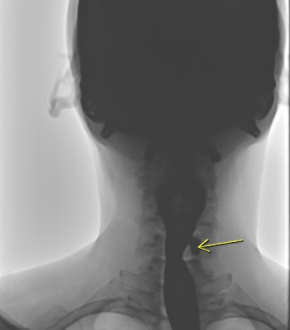
Many medical and surgical disciplines deal with KJD because of the wide range of symptoms and presentations. These include radiology, otolaryngology, gastrointestinal, and endocrinology. As a consequence of this, several forms of diagnostic imaging, such as CT scans, esophagoscopes, barium esophagram, and ultrasound, are utilized in the process of determining whether or not KJD is present.
Frequently, a barium esophagram is performed to diagnose a pharyngeal diverticulum. However, occasionally it could be challenging to differentiate ZD from KJD using only a barium esophagram, particularly if the diverticula are big. Therefore, another diagnostic technique such as a CT scan can be utilized to determine the diverticulum’s anatomical origin and to arrange the proper surgery.
Killian Jamieson Diverticulum Treatment
In the case of a symptomatic Killian-Jamieson diverticulum, traditional surgical therapy has been advocated due to the possibility of nerve harm. As a result of its reduced morbidity, lack of visible scarring, and speedier postoperative recovery, minimally invasive surgical techniques are increasingly popular now than ever before. Various endoscopic procedures are discussed, such as separating the intervening septum with a monopolar cautery or a carbon dioxide laser.
The anatomical link between the cricopharyngeal muscle and hypopharyngeal diverticulum is critical for treating Zenker’s diverticulum and Killian-Jamieson diverticulum. For Zenker’s diverticulum, surgery must include both diverticulectomy and cricopharyngeal myotomy. However, the Killian-Jamieson diverticulum does not need cricopharyngeal myotomy.
Killian Jamieson Diverticulum is treated endoscopically or surgically, depending on the severity of its symptoms or its size. Zenker’s diverticula that are less than 3 cm in diameter are often treated by endoscopic surgery. The diverticulum might become obstructed during endoscopic treatment if food or a foreign body is found there. In comparison to Zenker’s diverticulum, Killian Jamieson’s Diverticulum treatment is located closer to the recurrent laryngeal nerves. Other treatments, as opposed to endoscopic treatments, are preferred for KJD procedures that do not impact the recurrent nerves.


