The heart is the vital organ of the human body that is considered responsible for the provision of blood to all the organs for their proper functioning. This circulation of the heart is carried out in a well-arranged manner which involves the upward and downward flow resulting in the generation of some vibrations in the body. With every heartbeat, the blood is pushed into the blood vessels for further circulation, and these vessels are highly variable in size. At the point of variation of size, when blood enters into larger vessels like descending aorta than an upward twirl produces. This twirl is better described as a vibrational movement that can be observed at the surface of the body and responsible for the localized movement of that specified body part.
In general, these movements are called the ballistic forces characterized by the generation of repetitive 3D physical movements in the body resulting from the continuous flow of blood from the heart to the whole body. So, Ballistocardiography (BCG) is a non-intrusive procedure in which these ballistic forces are measured resulting from each heartbeat from the surface of the body. The reported frequency of these forces approximately falls in the range of 1 Hz to 20 Hz.
As a result of this technique a ballistocardiogram is obtained which is a representation of a person’s body recoil depending upon their heart rate and weight. This measurement is based on a BCG scale which provides the reference to weight and heart rate of a person for the accurate measurement of these ballistic forces. This data provides the manifestations of main heart dysfunctions and can be observed with a non-invasive procedure like non-contact cameras, which was firstly reported by Dr. Isaac Starr after a series of researches in 1940.
But in this initial era, the use of BCG to measure cardiac malfunctioning was very limited due to the complexity of the data obtained from this technique as well the large size of equipment used for this procedure. It required the use of some annotators which can further elaborate the data for the sake of understating. But over time the technology advanced and made the use of this technique easy and feasible for the professionals.
Ballistocardiography Sleep Monitor
Sleep apnea is characterized by the interruption or absence of external breathing which induces a certain disruptive pattern. This pattern can be detected by different techniques allowing the measurement of the Apnea-Hypopnea Index (IHA). Among other devices the measurement of BCG is considered helpful in this regard which is derived from a pressure modulated signal during the sleep apnea. BCG is known as a non-intrusive technique to measure the recoil of a human body generated by the cardiac pulsations.
It has been reported that the sleep apnea leads to different breathing patterns involving the affected cardiac patterns so that the measurement of these interrupted cardiac pulsations through BCG can help out to measure the AHI.
Emfit is a device made up of piezoelectric materials and designed for the sensing of pressure during sleep apnea. Various studies have shown the viability of accurately measuring BCG through these piezoelectric material-based devices. But regardless of the high sensitivity of these devices which is up to 0.95 can measure AHI <15, annotators are required to elaborate the picked up complex data.
Ballistocardiography Cardiac Output
Cardiac output is considered a very important factor to measure the proper functioning of the cardiovascular system. BCG is used prominently in this case and it typically lags ECG by the time of approximately 0.1 to 0.3 seconds and accurately measures the recoil of the body.
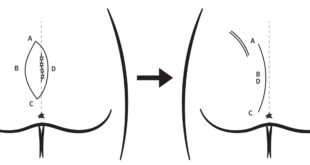
The obtained ballistocardiogram from this technique is based on a waveform which is further divided into three parts. The first part of the wave (GHI) describes the phase of before the ejection (expulsion) of the blood involving; venous return flow towards heart, filling of atria leading to contraction (G), upward refraction towards the head (H) and downward deflection towards lower limbs (I). The second part of the wave (J) represents the decreased flow in the head-ward area and hastening of blood flow in the abdominal portion of the body. Meanwhile, the third part of the wave represents the diastolic phase of the cardiac cycle.
Ballistocardiography Mechanism
The general mechanism of the BCG is based on the measurement of the vibrations generated by the repetitive flow of the blood from the heart to the whole body with each heartbeat. This flow generates vibrations of a certain frequency ranging from 1-20 Hz and can be measured with non-invasive devices made up of piezoelectric materials.
However, in the literature, three mechanisms are mentioned which are standardized and generalized in their nature but give out the different results of BCG. These can be differentiated based on their strength of frequency, including;
- Starr BCG (high-frequency BCG)
- Nickerson BCG (ultra-low frequency BCG)
- Dock BCG (direct-body BCG)
The advanced technology in the past few decades has helped a lot to regain the prominence of BCG.