A trichilemmal cyst is also known as a pilar, wen, or isthmus-catagen cyst. This cyst forms a hair follicle on the scalp most of the time and is visibly mobile, smooth, and filled with cytokeratin. Cytokeratin is a protein present in nails, hair, horns, and skin.
A trichilemmal cyst is different both clinically and histologically from trichilemmal horns. These horns are a rare hard tissue not restricted to the scalp. However, these cysts can grow more extensively and multiply rapidly as trichilemmal tumors. For this reason, trichilemmal cysts are known as proliferating trichilemmal cysts that are benign but may grow at the cyst site aggressively. It happens very rarely that these cysts can become cancerous. Although the trichilemmal cysts or pilar cysts are not necessarily a cause to be bothered about, you may find their growth uncomfortable. A person may identify them but, it is better to consult a doctor for an official diagnosis.
Trichilemmal Cyst Symptoms
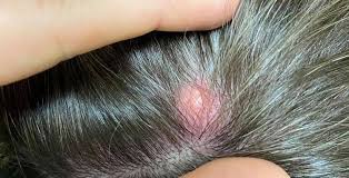
90% of the trichilemmal cysts are present on the scalp; they can develop anywhere on the body. Other possible sites of their occurrence include the neck and face. Most of the time, affected individuals may end up having more than a single cyst. These cysts appear as hair follicles which is a characteristic feature of them.
These cysts can range from the size of a quarter to the size of a ball as time passes. These are the same as your skin color. Trichilemmal cysts sometimes create a dome-like bump on the skin surface. These cysts are firm to the touch but smooth in texture. One characteristic of these cysts is that they are not painful and usually do not contain pus. A cyst sometimes ruptures due to trauma or on its own. In case if the rapture takes place, pain, rash or irritation in the affected area is noticeable.
Trichilemmal Cyst Causes
Trichilemmal cysts appear when the epidermis sheds old skin cells. These shedding cells seldom shift under the skin surface instead of shedding off. When this shifting of the shedding cells occurs, the shredded cells begin to multiply as if they are still in the process of growth. Rather than moving up to the surface, they stay trapped under the skin and start building up.
Meanwhile, the scalp releases keratin responsible for the maintenance of hair health.
The keratin builds up with the dead cells of the skin. As a result, a white-yellow liquid oozes out from the cyst if it ruptures. If the pilar cyst ruptures, the affected individuals may also be at an increased risk for swelling and irritation at its site.
With time, the protein in the follicle creates a bump, which is a characteristic of a pilar cyst. These cysts may be hereditary and are more frequent in middle-aged women.
Trichilemmal Cyst Diagnosis
The diagnostic method of the trichilemmal cyst involves a simple visual examination. As these cysts can resemble other skin conditions, for this reason, the doctor will need to figure out other health concerns. He may ask you questions about the symptoms. A biopsy of the pilar cyst can also aid to diagnose this benign cyst.
A small tissue sample of the affected area is taken and sent to the lab for microscopic evaluation.
A person may self-diagnose this cyst based on visible risk factors and signs. However, it is necessary to consult a doctor to confirm this condition. The doctor will rule out underlying causes that may be even more complicated. Sometimes a CT scan is also performed to eliminate cancer and other types of cysts. The abovementioned diagnostic strategies can also give information about the underlying layers of the cysts to aid in seeing if any more cysts are forming.
Trichilemmal Cyst Treatment
The treatment of a trichilemmal cyst involves surgical excision. However, the treatment method varies depending upon the physician’s training. The majority of the physicians perform the surgery by using a local anesthetic. The other approach is more conservative and involves a small punch biopsy about one-fourth the cyst’s diameter. This biopsy enters the cyst cavity. The cyst contents are removed and an empty sac is left behind. The pillar cyst wall is the most durable and thickest variety among other varieties of cysts. It is grabbed by using forceps and pulled out of the incision. This method is suited best on larger cysts with a well-formed thick wall so that the identification becomes clear.
However, the small cysts are thin-walled; these are readily fragmented; on traction. It increases the likelihood of cyst recurrence. This method often results in a small scar and, very little, if any bleeding, is evident.


