A surgical procedure called a subxiphoid pericardial window tells if there is blood in the sac surrounding the heart. The goal is to incise the pericardium under direct visualization to check for blood in the pericardial sac, which is only done surgically. It is most commonly utilized as a diagnostic technique in cases of penetrating damage, but it is also necessary in cases of blunt trauma. When treating symptomatic pericardial effusion, a subxiphoid pericardial window is typically recommended as the treatment.
Medicines are often employed to minister pericardial effusion, depending on the degree of accumulation. If the healthcare team believes that it is important to drain the fluid buildup, they often suggest a treatment called pericardiocentesis, which involves the use of a needle and a tiny catheter to drain the fluid. This procedure is only performed if it is determined that it is required to drain the excess fluid.
When treating symptomatic pericardial effusion, a subxiphoid pericardial window is typically recommended as the treatment.
To execute a subxiphoid pericardial window, either general anesthesia or local anesthetic with sedation is used, with the latter option reserved for hemodynamically stable patients. It is carried out within an operating room.
Subxiphoid Pericardial Window Procedure
Subxiphoid pericardial draining is best done under general anesthesia, however severe tamponade patients who do not tolerate it prefer it done under local anesthetic and sedation. If general anesthesia is utilized, the skin of the patient is prepped and draped before induction to avoid a delay in the event of hypotension.
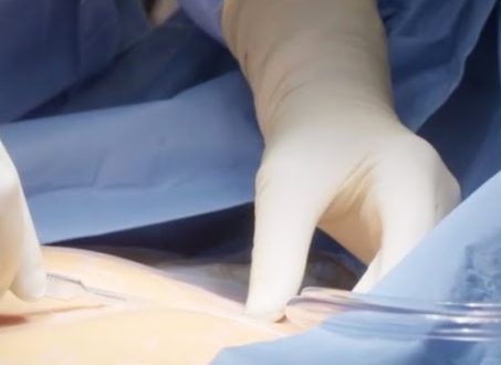
It is necessary to make a short incision that extends caudally from the bottom of the sternum for about 6 to 8 centimeters. The xiphoid sternum is resected and the upper linea alba is separated at its midpoint. The peritoneum remains closed. By using a blunt finger dissection technique, the tissue plane between the anterior pericardium and the posterior wall of the sternum is formed.
A small retractor is inserted into the abdominal incision, and the distal sternum is lifted with a right-angled retractor. After making an incision in the anterior pericardium, the fluid is drained and then sent for bacteriological and cytologic examination.
A digital examination of the pericardium is carried out to locate any adhesions or tumor deposits. Bacteriological and histologic examinations of a pericardial biopsy sample that is 2 to 3 centimeters in diameter are performed. Postoperative suction drainage is accomplished by inserting a 28 F chest tube through the pericardiotomy, which is accessed through a second stab hole in the upper left abdomen.
Subxiphoid Pericardial Window Ultrasound
The traditional pericardial tamponade symptoms, such as raised central venous pressure, muted heartbeats, and paradoxical pulse, are useless in an emergency. The diagnostic usefulness of electrocardiograms and chest roentgenograms is minimal.
In most cases, diagnostic imaging is performed, such as an echocardiography or chest CT scan. Before undergoing surgery, it is essential to examine these investigations to gauge the extent of the effusion and establish whether the effusion is located primarily in the anterior or posterior compartments. When using the subxiphoid technique, the surgeon spends the majority of their time gaining access to the pericardium anteriorly, which is over the right ventricle.
Subxiphoid Pericardial Window Technique
This surgery is conducted through an incision in the upper midline of the abdomen. Retracting the xiphoid process or removing it entirely are both possible options. The tiniest part of the sternum, often known as the breastbone, is called the xiphoid process. When a baby is first born, it is formed of cartilage, but as an adult, it turns into bone.
Utilizing a “Rultract retractor” is the most recommended method. In addition to liberating the surgical assistant, this allows for great pericardial and substernal region exposure. The treatment is conducted under either general or local anesthetic. It is performed reasonably quickly, immediately alleviates symptoms, and permits direct digital examination of the pericardial sac. A big-diameter drainage tube is installed to facilitate drainage and suction if required.


