Learn all about staghorn calculus definition, symptoms, causes and treatment.
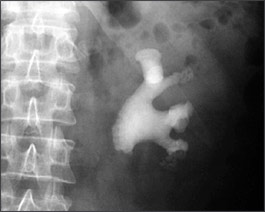
Staghorn calculi represent a less-common nephrolithiasis subgroup so named because the significant stone burden that fills the renal pelvis and calyces forms a shape on radiographs that resembles a deer’s horns. Most staghorn stones in Western society are composed of struvite and can cause significant morbidity and mortality if left untreated; therefore, large struvite stones must typically be removed.
Staghorn stones are large, branched stones occupying a large volume of the kidneys. The most effective treatment option is percutaneous nephrolithotomy, where the urologist inserts a scope directly into the kidney via a 1/2 inch tract made in your flank. Open surgery for kidney stones is strongly discouraged and reserved for only very rare cases with a very large stone burden or very unusual anatomy.
Staghorn stones are stones that involve the renal pelvis and extend into at least 2 calyces of the kidney. Approximately 75% of staghorn calculi are struvite (composed of magnesium, ammonium, phosphate crystals mixed with carbonate-apatite). These stones are associated with urinary tract infections caused by bacteria that produce urease. Urease hydrolyzes (or splits) urea into ammonium and hydroxyl ions resulting in an increase in ammonium and phosphate concentrations leading to an alkaline urine.
The alkaline urine leads to precipitation of magnesium ammonium phosphate crystals mixed with varying proportions of carbonate apatite. The infection stones generally grow rapidly and serve as the nidus (or center) for further stone formation. This stone matrix is believed to protect the bacteria from antibiotics. In essence, antibiotics cannot clear the associated infection unless the stone material is completely removed.
What is Staghorn Calculus?
Staghorn calculi refer to branched stones that fill all or part of the renal pelvis and branch into several or all of the calyces. They are most often composed of struvite (magnesium ammonium phosphate) and/or calcium carbonate apatite. These stones are often referred to as ‘infection stones’ since they are strongly associated with urinary tract infections with urea splitting organisms. Cystine stones, although much less common, may also develop into staghorn calculi.
Staghorn Calculus Definition
Staghorn calculi, also sometimes called coral calculi, are renal calculi that obtain their characteristic shape by forming a cast of the renal pelvis and calyces, thus resembling the horns of a stag.
Staghorn Calculus Symptoms
Symptoms may include flank pain classic for renal colic, fever, urinary symptoms (eg, frequency, dysuria), and hematuria (either gross or microscopic). 25% of patients may remain completely free of symptoms.
- Severe pain in the side and back, below the ribs.
- Pain that radiates to the lower abdomen and groin
- Pain that comes in waves and fluctuates in intensity
- Pain on urination
- Pink, red or brown urine
- Cloudy or foul-smelling urine
- Nausea and vomiting
- Persistent need to urinate
- Urinating more often than usual
- Fever and chills if an infection is present
- Urinating small amount
Staghorn Calculus Causes
Staghorn calculus can be caused due to various types of stones.
Calcium stones: Most kidney stones are calcium stones, usually in the form of calcium oxalate. Oxalate is a naturally occurring substance found in food and is also made daily by your liver. Some fruits and vegetables, as well as nuts and chocolate, have high oxalate content.
Dietary factors, high doses of vitamin D, intestinal bypass surgery and several metabolic disorders can increase the concentration of calcium or oxalate in urine. Calcium stones may also occur in the form of calcium phosphate. This type of stone is more common in metabolic conditions, such as renal tubular acidosis. It may also be associated with certain migraine headaches or with taking certain seizure medications, such as topiramate (Topamax).
Struvite stones
Struvite stones form in response to an infection, such as a urinary tract infection. These stones can grow quickly and become quite large, sometimes with few symptoms or little warning.
Uric acid stones: Uric acid stones can form in people who don’t drink enough fluids or who lose too much fluid, those who eat a high-protein diet, and those who have gout. Certain genetic factors also may increase your risk of uric acid stones.
Cystine stones
These stones form in people with a hereditary disorder that causes the kidneys to excrete too much of certain amino acids (cystinuria).
Staghorn Calculus Treatment
Staghorn calculus treatment can be done both medically and surgically. A guideline from the American Urological Association recommends that patients with residual or recurrent struvite stones may be offered treatment with acetohydroxamic acid (AHA), but only after surgical options have been exhausted. Suppressive antibiotic therapy may prevent pyelonephritis and associated systemic infection and may help inhibit stone growth.
Other medical interventions, such as urinary acidification with oral ammonium chloride, have had limited long-term clinical utility. The advent of minimally invasive modalities to treat renal stones revolutionized the approach to staghorn calculi. These include extracorporeal shockwave lithotripsy (SWL) and percutaneous nephrolithotomy (PNL).


