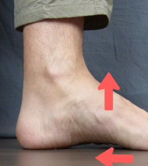
The hindfoot is the portion of the foot that extends from below the ankle to above the Chopart joint. Hindfoot valgus is characterized by a displacement of the mid-calcaneal line from the midline of the body. From a dorsoplantar vantage point, this results in a greater angle between the mid-calcaneal and mid-talar axes. The mid-calcaneal line does not change much when the heel bone goes forward. Sometimes it passes over the third metatarsal base instead of the fourth, for example, indicating a more medial crossing of the metatarsal bases. The talus appears medially angulated due to the widening of the talocalcaneal angle, with the mid-talar axis reaching all the way to the first metatarsal’s base.
Flatfoot is characterized clinically by a flattening of the medial longitudinal arch and valgus malformation of the hindfoot. Physiological flatfoot, in which the foot is contracted but still pliable, develops as a congenital or acquired deformity and is considered in the differential diagnosis of flatfoot. Congenital flatfoot deformity needs intensive treatment right away, but a flexible flatfoot in a child has a good outlook, and conservative treatment frequently leads to a stable foot that often carries enough weight.
Hindfoot Valgus Symptoms
Most frequently, middle-aged and older people with persistent hindfoot valgus deformity experience posterior hindfoot impingement. Limited subtalar ranges of motion and hindfoot pain with weight bearing are common symptoms, as well as edema and tenderness in the area anterior and posterior to the lateral malleolus. These symptoms are not particular and occur in patients with other conditions affecting the hindfoot.
Hindfoot valgus deformity is frequently brought on by posterior tibial tendinopathy. Higher degrees of posterior tibial tendon rupture have been observed to enhance the occurrence of lateral hindfoot impingement. Patients with the malfunction of the posterior tibial tendon feel both discomfort and impairment. Initial foot discomfort is felt along the medial side and is frequently accompanied by tenosynovitis-related edema.
Hindfoot Valgus Causes
The presence of a valgus malalignment in the hindfoot is essential for the development of lateral hindfoot impingement. Anatomical changes also make it difficult to get measurements in the same place every time.
Inflammatory arthropathies, such as rheumatoid arthritis and psoriatic arthropathy, are associated with an increased risk of tibialis posterior tendon tear due to chronic inflammation at the tendon sheath and neighboring joints, leading to an increased prevalence of hindfoot valgus collapse. One of the most common causes of hindfoot valgus deformity is a condition known as posterior tibial tendinopathy.
It is discovered that more severe cases of posterior tibial tendon tear are associated with a higher incidence of lateral hindfoot impingement. Pain and limited mobility are common complaints among those who suffer from dysfunction of the posterior tibial tendon. The discomfort is initially felt along the medial part of the foot, and it is frequently coupled with swelling caused by tenosynovitis. With the gradual collapse of the longitudinal arch and the development of a valgus deformity in the back of the foot, lateral foot pain develops. This pain is often caused by talocalcaneal or calcaneofibular impingement, which happens outside of the joint.
Hindfoot Valgus Exercises
In individuals with symptomatic flatfoot, which is typically caused by tendon insufficiency of the tibialis posterior, conservative treatment with insoles, shoe adjustments, and physiotherapeutic techniques often lead to significant improvement; otherwise, surgical correction is recommended. Early therapy that is right for the child’s age helps stop the foot from getting worse.
Intramuscular lengthening is the method of choice because it reduces the strength of the peroneus brevis by one point on the MRC scale and corrects this condition by one grade of severity. Tendon lengthening is most effective when used on mild to moderate deformities.
Hindfoot Valgus Surgery
Arthritis and deformities of the midfoot and hindfoot mostly result in incapacitating discomfort and functional impairment. Unlike some other joints in the lower body, there are not many surgical options for the affected joints other than arthrodesis. Alterations to footwear and routine, as well as the use of orthotics, often form the basis of initial treatment.
Surgery offers the potential to treat both the valgus deformity and the osteoarthritis that develops in the knee joint as a result of the passage of time. In patients who are still relatively young, an osteotomy surgical treatment is a viable alternative. To reposition the knee and rectify the placement, the femur (thigh bone) is sliced.
 Health & Care Information


