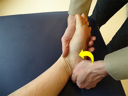
Chronic medial heel pain may be a common complaint in running-based athletes. In most cases, the source of pain is possibly plantar fasciitis. However, another potential source of pain is the first branch of the lateral plantar nerve, referred to as Baxter’s nerve. Often, Baxter nerve neuropathy can coexist with plantar fasciitis.
Baxter’s neuropathy is a pinched nerve syndrome that results from compression of the lower calcaneal nerve. Causes of Baxter neuropathy include changes within the biomechanics of the foot like flat feet, plantar calcaneal enthesophytes, and plantar fasciitis.
What is Baxter’s Neuropathy?
Baxter’s entrapment is an entrapment (or compression) of the Inferior Calcaneal Nerve slightly below the foot’s arch. The Inferior Calcaneal Nerve is the first branch of the Lateral Plantar Nerve on the foot’s underside. The nerve is additionally called Baxter’s nerve, named after the primary physician, to explain this nerve compression as a selected explanation for foot pain.
Baxter’s Neuropathy Symptoms
Below are a few signs and symptoms of Baxter nerve entrapment that can help a doctor distinguish this problem from plantar fasciitis.
● The pain is usually more proximal and medial, usually distal to the medial tubercle of the calcaneus.
● No early morning pain, instead of the problem that tends to get worse during the day, or pain after prolonged activity.
● When the area of the nerve is touched by the hand, radiating pain may appear.
● The pain increases with passive eversion and abduction of the foot.
● Presence of maximum soreness in the middle of the heel where entrapment occurs. This can cause radiating and burning pain across the plantar foot.
● Baxter’s Nerve Entrapment is may be possible due to the low abduction of the fifth toe. It is related to ADMM.
● In chronic cases, patients may have decreased sensitivity of the lateral plantar foot.
● There may be a positive Tinel sign. Paresthesias can be reproduced by tapping a nerve under the abductor’s muscle.
Baxter’s Neuropathy Causes
There are several factors that can predispose a person to Baxter neuropathy; this includes:
● Obesity
● A sudden increase in running distance
● Jobs that require long durations of standing or walking
● Flat foot
● Plantar fasciitis
● Limited range motion of the ankle
● Muscular Enlargement (such as in athletes)
All of these factors, in one way or another, affect the likelihood of Baxter neuropathy. They alter the foot’s biometric characteristics and thus increase the tensile and compressive forces in all structures.
Baxter’s Neuropathy Treatment
Treatment for a compressed Baxter nerve is similar to the initial treatment for plantar fasciitis. The following methods can be used initially:
● Tape and orthopedic devices to control pronation
● Stretching the soleus and calf muscles
● Therapy for the soft tissue of the plantar fascia and inner foot
● Strengthening exercises for the internal organs of the foot
Patients with an entrapped Baxter nerve often have a relatively slow and low response to conservative treatment. Therefore, if symptoms persist for more than six months, the next course of action is to inject a steroid / local anesthetic into and around the nerve. It is both diagnostic and therapeutic.
If symptoms improve after injection, this indicates a pinched nerve that is the source of the symptoms. Otherwise, plantar fascia injury is the most likely scenario. If local steroid injection fails, surgery may be required. This can be endoscopic nerve decompression, radiofrequency ablation, or open surgery.
Physical Test for Baxter’s Nerve Entrapment
A physical test that a clinician can use is an adaptation of the tibial nerve test. Since the lateral plantar nerve branches off from the tibial nerve, a tibial nerve test can be used to identify a neurodynamic interface problem with Baxter’s nerve. This test is carried out as follows:
The patient lies on his back. The doctor holds the affected foot and pushes the foot into dorsiflexion of the ankle and notes the symptoms. The physician should then raise the leg to flex the hip, with the knee in extension and the foot in dorsiflexion. Pay attention to any signs, especially pain in the middle of the heel. To determine if the problem is with a nerve, a tendon problem, or something else, you need a sensitizer that changes the tension on the nerve. To do this, slowly lower the hip out of flexion and note whether the symptoms have changed. If the pain in the mid-heel is relieved with less hip flexion, the tibial nerve and its branches are affected.
 Health & Care Information


