Learn all about ureterocele and its causes, symptoms and treatment options. Ureterocele is a dilation of the area where the ureter inserts into the bladder. Ureteroceles are a congenital abnormality which means they are present at birth. It is unknown exactly why ureteroceles form, but it is thought to be due to an abnormality of the formation of the ureter tube as it inserts into the bladder.
If the ureterocele is associated with a duplex kidney, it is the ureter that drains the upper part of the kidney that has the ureterocele. Kidneys that have ureterocele often are dilated and can have obstruction to urine flow.
Ureteroceles occur in approximately 1 in every 4000 children and occur most commonly in whites. Females are affected 4-7 times more often than males. In the adult population, ureteroceles also occur more frequently in females. Orthotropic ureteroceles occur in 17-35% of cases, with an incidence of ectopic ureteroceles of approximately 80% in most pediatric series. Similarly, approximately 80% of ureteroceles are associated with the upper pole moiety of a duplex system.
Ureteroceles may be categorized based on their relationship with the renal unit or based on distal ureteral configuration and location. The main problem from ureterocele is kidney damage, and kidney infection. Urine blockage may damage the developing kidneys and reduce their ability to filter.
No single approach is appropriate for all patients with ureteroceles; therefore, each case must be tailored to the individual. An experienced surgeon must be armed with various surgical techniques that can be tailored to effectively treat different types of ureterocele malformations.
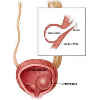
What is Ureterocele
An ureterocele is a cystic out-pouching of the distal ureter into the urinary bladder. Ureterocele is a swelling at the bottom of one of the tubes (ureters) that carry urine from the kidney to the bladder. The ureterocele is a birth defect.
Ureterocele Pictures
Ureterocele Symptoms
Ureteroceles may be asymptomatic or may produce a wide range of clinical signs and symptoms, from recurrent cystitis to bladder outlet obstruction to renal failure. Because of the obstructive nature of ureteroceles, the activity of the affected renal unit varies from a normal, well-functioning kidney to a nonfunctioning, dysplastic renal segment or kidney. However, with proper diagnosis and treatment, the outcome remains excellent. Ureteroceles often do not have any symptoms, unless the condition has resulted in a urinary tract infection (UTI). Other symptoms of ureterocele include:
· painful urination
· bad-smelling urine
· blood in the urine
· fever
· urinating often
· burning feeing while urinating
· a lump in the abdomen
· urinary incontinence (inability to hold urine)
· not being able to empty the bladder
· Cyclic abdominal pain
· Urolithiasis
· Cobra head sign is seen in radiography
· salpingitis, hydrosalpinx with sepsis or torsion
Ureterocele Causes
A ureterocele occurs during the prenatal period when the ends of ureters that enter the bladder don’t develop properly. Though a ureterocele is considered a birth defect, its etiology is still unknown. It has been suggested that genetics might play a role. The condition runs in families, but to date no specific genes have been isolated. A ureterocele occurs in the lower part of the ureter, where the tube enters the bladder. The swollen area prevents urine from moving freely into the bladder. The urine collects in the ureter and stretches its walls. It expands like a water balloon. However, certain situations may cause the pelvic floor muscles and tissues to lose their natural strength, causing them to stretch and become unable to hold the urethra (other structures of the female organ) in their original place.
Ureterocele Diagnosis
Many children with ureteroceles are identified during fetal ultrasounds performed before birth, but they may not be diagnosed until a patient is being evaluated for another condition such as a urinary tract infection
Ultrasonography is used to allow radiologists to diagnose a ureterocele. A bladder test, the voiding cystourethrogram (VCUG), is usually performed to define the type of ureterocele, the effect on bladder function and if there is any reflux into any of the ureters. Renal scan (DMSA or MAG-3) is often performed to evaluate the function of the kidneys, specifically to determine whether the affected portion of the kidney has any function. Abdominal CT scans and MRI tests are additional studies that may also be performed in situations where the urinary tract anatomy is extremely ambiguous and will allow the surgeon to better identify anatomical variations.
Ureterocele Treatment
Ureterocele treatment includes the followings;
- Surgery.
- Transurethral Puncture.
- Upper Pole Nephrectomy, this operation is done either through a small cut under the ribs, or laparoscopically.
- Nephrectomy.
- Removal of the Ureterocele and Ureteral Reimplantation.
- Ureteropyelostomy or Upper-to-Lower Ureteroureterostomy; the operation is done with a small incision in the lower abdomen. The success rate is 95%.
- Antibiotics are used to fight bacteria and prevent kidney infection. A child with a possible urine block or urine reflux may be given antibiotics to prevent infections until the defect is corrected.
Ureterocele Ultrasound
Focused ultrasound could potentially treat ureteroceles because of its ability to non-invasively destroy target tissue through cavitation (histotripsy). However, research is still in the early pre-clinical stages. In a recent feasibility study, scientists at the University of Washington were able to use successive pulses of focused ultrasound to achieve tissue fractionation (punctures) in a tissue model of a ureterocele wall. On antenatal sonograms, the typical presentation is that of an enlarged hydronephrotic fetal kidney. Renal duplication and/or ureteral duplication may be seen. Intravesical cystic dilation or septa within the bladder (which represent ureterocele walls) suggest that the etiology of the hydronephrosis may be a ureterocele. Such antenatal ultrasonographic findings warrant comprehensive postnatal evaluation and confirmation of the diagnosis.


