Learn all about arachnoid granulations. It is named after Antonio Pacchioni, an Italian anatomist (1665-1726), who wrote extensively on the anatomy of the dura mater, published in 1705.
Arachnoid granulations may expand the dural sinuses or inner table of the skull. Although usually incidental, giant arachnoid granulations that are of sufficient size to fill the lumen of a dural sinus and cause local dilation or filling defects can rarely cause symptoms due to sinus obstruction leading to venous hypertension.
Arachnoid granulations normally measure a few millimeters. In some cases they grow to fill and dilate the dural sinuses even with expansion of the inner table of the skull and are then called “giant AG” 1. There is no consensus on the definition of “giant” in the literature. Sometimes large arachnoid granulations are called “giant” when larger than 1 cm. Most commonly AG is found in the transverse sinuses, particularly within the middle and lateral portions of the sinus, with a slight left predominance 6. The second most common location is the superior sagittal sinus, but they can be found everywhere in the dural venous sinuses 2, 4. There is no difference in sex distribution.
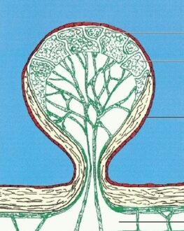
The arachnoid villi absorb excess cerebrospinal fluid, or CSF, that collects in the venous sinuses surrounding the brain. Acting as one-way valves, the arachnoid villi, or arachnoid granulations, help to ensure that the pressure and volume of CSF surrounding the brain does not reach dangerous levels.
The arachnoid granulations often have a parasagittal location, leading to specific changes on the X-rays of the skull, such as the osteolytic lucency or the filling defect that appears at the level of the dural venous sinuses. Because of the latter modification, it is possible to mistake the arachnoid granulation for dural venous thrombosis. The MRI can be used for the identification of these small protrusions, with general signal characteristics for the cerebrospinal fluid.
What is Arachnoid granulations
Arachnoid granulations, also known as a Pacchionian granulation, are projections of the arachnoid membrane (villi) into the dural sinuses that allow CSF entrance from the subarachnoid space into the venous system. These are small protrusions of the arachnoid mater (the thin second layer covering the brain) through the dura mater (the thick outer layer). They protrude into the venous sinuses of the brain, and allow cerebrospinal fluid (CSF) to exit the subarachnoid space and enter the blood stream.
Arachnoid granulations Symptoms
The clinical significance of arachnoid granulations is uncertain. Most cases are usually asymptomatic and there are incidental findings on imaging studies, while some giant AGs may cause dural venous sinus pressure gradients and headache. In this case, the headache might be closely associated with multiple AGs. Imaging of giant arachnoid granulations is often suggestive of partial sinus occlusion.
Although largely incidental, giant arachnoid granulations can rarely cause symptoms due to sinus obstruction leading to venous hypertension. Dural sinus pressure measurement across the lesion is a novel and valuable approach to determine if the lesion is symptomatic because of venous obstruction and hypertension. Normal venous pressure with no significant differential pressure across the lesion can safely exclude the lesion as the cause of the patient’s symptoms.
Arachnoid granulations Causes
The causes associated with arachnoid granulations are as under;
- Aqueductal stenosis
- Neural tube defect
- Arachnoid cysts
- Dandy-Walker syndrome it may also be associated with abnormal development in other parts of the brain and sometimes leads to aqueductal stenosis.
- There are two types of Chiari malformation. Both types occur in the bottom of the brain stem where the brain and spinal cord join. The lowest portion of the brain is displaced and is lower than normal pushing down into the spinal column.
- An intraventricular hemorrhage, which most frequently affects premature newborns, may cause an acquired form of hydrocephalus. When small blood vessels alongside the ventricular lining rupture, blood may block or scar the ventricles or plug the arachnoid villi, which allow CSF to be absorbed. When the CSF can’t be absorbed, hydrocephalus results.
- Meningitis
- Head injury
Arachnoid granulations Treatment
Arachnoid granulations treatment has been active debate about how to treat this condition. The need for treatment depends mostly upon the location and size of the villi whether it is small not disturbing surrounding tissue, and not causing symptoms, some doctors will refrain from treatment. Now with micro neurosurgical techniques and endoscopic tools that allow for minimally invasive surgery, more doctors are opting to surgically remove the membranes of the granulations or open them so its fluid can drain into the cerebrospinal fluid and be absorbed.
A patient with pseudo tumor cerebri syndrome who’s venography showed a giant arachnoid granulation in the left dominant transverse sinus and hypoplasia of the contralateral transverse sinus with high pressure proximal to the obstruction. Dilation of the left transverse sinuses with a stent reduced both the pressure gradient across the arachnoid granulation and the cerebral spinal fluid opening pressure with immediate symptomatic improvement, suggesting a causal relationship between venous outflow obstruction and pseudo tumor cerebri syndrome.