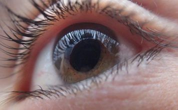
Learn all about dyschromatopsia definition, test, causes, symptoms and treatment. Dyschromatopsia is a condition in which the person is not able to distinguish between different colors.
The causes that cause this disease are diverse. Individual from birth may have a sensitivity to certain colors but it is also possible that the appearance of cataracts throughout life to accelerate or trigger dyschromatopsia. In addition, from a simple myopia that could increase with age until the appearance of this disease suddenly appears to be only a step.
Some medications or diseases (pernicious anemia or sickle-shaped cell disease) can also trigger the disease. It seems that the blame for this situation are cones, specialized cells in the retina, light wave essential for translating colors perceptible. These cells are completely absent from birth, may be missing part or may not work well, leading to this lack of perception of color.
The fact that the individual could pass the Ishihara test using this lens has led us to believe that there may be a case for exploring this therapeutic option further. This may potentially aid in functional rehabilitation of those personnel who are already trained and have joined service based on an erroneous initial medical evaluation and are later detected to have substandard colour perception not meeting the occupational requirements.
Dyschromatopsia can be the initial symptom in toxic/nutritional optic. Amiodarone is a drug very useful in the treatment of life-threatening. This case highlights the importance of a thorough ophthalmic examination when dealing with candidates who will make every effort to gain entry into the armed forces. Ophthalmic evaluation under good illumination and magnification is a must at the level of initial examination to avoid any errors.
What is Dyschromatopsia?
Some people do not distinguish red from green, or confuse others combine different colors and you can see only in a certain light, and in rare cases do not distinguish colors at all. This lack of perception of color is called dyschromatopsia.
Dyschromatopsia Definition
A condition in which the ability to perceive colors is not fully normal. Dyschromatopsia are alterations of our color vision. A congenital defect or an acquired defect in discriminating one colour from another
Dyschromatopsia Test
The color vision test for diagnosis and evaluation of the dyschromatopsia consists of a set of 92 colored test charts of the pseudo chromatic type, a lamp and a test report giving a graphic image of the sensitivity to color deficiency. This test was created in 1917 by Ishihara Shinobu.
- It is formed by 38 plates called “pseudo isochromatic”, plates made of little colored circles, forming a background on which appears a symbol (letter or number). Patients who do not have any dyschromatopsia clearly distinguish the symbol, while those who do suffer from the condition will have difficult. Its purpose is to highlight the existence of a potential dyschromatopsia, but don’t enable the detection of its seriousness.
- In the Holmgren test, the patient has to select a certain number of similar colors from amongst a bunch of colored threads.
- In the Farnsworth test, the patient has to group or put in order some coloured tokens.
Dyschromatopsia Causes
The dyschromatopsia are caused by mutations affecting genes encoding pigments responsible for color vision. The blue-sensitive pigment is on chromosome 7. Pigments sensitive to red and green are encoded in a gene on the X chromosome, so its inheritance is sex-linked. The mutations are recessive, which means that only manifest if both copies of the gene are altered. For a woman to suffer alterations in their view of the red or green you must have the corresponding gene mutated in their two X chromosomes; while men, to have a single X chromosome, they suffer alteration if any X chromosome inherited the lead. This explains that the frequency of these abnormalities is greater in males than in females.
Dyschromatopsia Symptoms
The major symptoms of dyschromatopsia are vision loss, usually in one eye, often developing within hours to a couple of days and peaking in one to two weeks. It may vary from a small area of blurring to complete blindness. The five symptoms associated with dyschromatopsia are:
- Achromatopsia.
- Amblyopia (reduced visual acuity)
- Hemeralopia (with the subject exhibiting photophobia)
- Nystagmus.
- Iris operating abnormalities.
Dyschromatopsia Treatment
Dyschromatopsia treatment is done by medications or lens. The antituberculous drug ethambutol is commonly associated with toxic optic neuropathy (not optic neuritis). This is the drug’s most serious adverse effect. The optic neuropathy that occurs is dose dependent and duration related. The chelating properties of ethambutol have been hypothesized to contribute to its neurotoxicity, causing calcium flux into the mitochondria. Although there is no evidence that the use of oral nonsteroidal anti-inflammatory drugs (NSAIDs) have an effect on ultimate visual outcomes, they are effective in decreasing the pain on eye movement often associated with optic neuritis. Various disease-modifying agents, including Glatiramer acetate (Copaxone), interferon beta-1a (Avonex, Rebif), interferon beta-1b (Betaseron, Extavia), mitoxantrone (Novantrone), and natalizumab (Tysabri), are used to decrease episodes of demyelination in patients with suspected or proven multiple sclerosis.
 Health & Care Information


