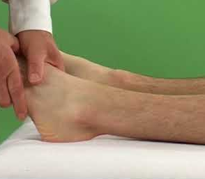
The foot and the leg are connected at the ankle, which functions as a hinge joint that allows for movement in both directions (up and down). Talus, the third and final foot bone, slides into a slot formed by the tibia and fibula. Ligaments are responsible for connecting the talus to the tibia and the fibula.
The term “high ankle ligament” is frequently used to describe the anterior tibiofibular ligament. This ligament slopes distally and laterally downward between the fibula and tibia margins. The ligament connects the fibula and tibia bones just above the ankle joint (syndesmosis). Walking and running put a lot of pressure on the tibia and fibula, so keeping them stable is essential. A sprain of the ankle ligaments is much more common than an injury to the upper ankle joint.
Anterior talofibular ligament Function
The Anterior Talofibular Ligament’s job is to keep the ankle joint from turning inward and bending down toward the ground. Injuries to the Anterior talofibular ligament are common in athletes whose centers of gravity are shifted laterally over the weight-bearing leg, leading to rapid inward rolling of the ankle. The anterior talofibular ligament is the most vulnerable of the lateral collateral ligaments, and as a result, it is the one that usually gets injured first. This ligament is crucial in preventing the talus from moving forward and the ankle from flexing in the plantar direction. This ligament is typically made up of two bands, and it is closely connected to the joint capsule of the ankle.
Anterior talofibular ligament Tear
Sprain injuries, which limit the motion of the ankle, happen when an ankle ligament is bruised, stretched, or torn. When the foot is planted unnaturally or when the ankle is twisted awkwardly, the anterior talofibular ligament, which is located on the lateral side of the ankle, is responsible for absorbing the majority of the negative impact. However, if the ligament is partially or entirely torn, the damage is significantly worse. A mild anterior talofibular ligament strain heals in three to four days. Ankle inversion injuries most frequently involve the anterior talofibular ligament, which is the weakest of the lateral collateral ligament complex and one of the lateral stabilizing ligaments of the ankle.
Ankle sprains often lead to instability in the ankle. The most common reason for ankle instability is a tear in the anterior talofibular ligament, also known as the ATFL. Arthroscopic ankle instability treatment is a new field that is gaining popularity among surgeons. During an arthroscopic all-inside ATFL repair, the surgeon is able to examine the ankle joint and reattach the torn ATFL to its anatomical location in the fibula.
Anterior talofibular ligament Pain
Inversion causes stress on the ankle ligaments and often results in injury. An extreme ankle sprain occurs when the anterior tibiofibular ligament is overstretched or torn as a result of the body’s weight being placed on the lateral ankle ligaments. Sometimes, the ligament is also torn through bone fragments. This ankle-twisting force occasionally results in additional harm. There is a high risk of injury to the bones, cartilage, ligaments, and tendons in and around the ankle joint.
Syndesmosis injuries are the most severe type of ankle and foot sprains, and they cause many complications for people who are trying to return to their normal activities. Most often, when an ankle is sprained, the affected side experiences sharp, localized pain. Ankle pain is the first symptom experienced by many people with mild or moderate syndesmosis sprains. Pain and swelling on the outside of the ankle are common symptoms, as are skin discoloration and bruising. The ankle feels shaky and weak, and pain mostly radiates along the side of the lower leg.
Anterior talofibular ligament Repair Surgery
During surgery, the damaged ATFL ligament is typically tightened and reattached to the bone so that it regains its original shape and strength. The replacement of a damaged ligament with a tendon graft becomes necessary if the ligament is too weak to be repaired.
The patient is positioned supine with their lower extremities rotated slightly inward and slightly elevated. A skin incision is made using a minimally invasive open technique, beginning 2 cm from the fibula’s apex along its posterior rim, running parallel to the foot, and roughly following the location of the former ligament. The incision measures about 5 cm in length. The damaged ligament pieces are located.
 Health & Care Information


