Bones are the strong and main pillars of the body but, when lumps of abnormal tissues are formed and, uncontrollably, cells divide within a bone, then bone tumors are developed and make them weak. Fibrous Cortical is also a bone tumor, which occurs most frequently in children and young adults. This defect heals naturally as a child’s bone stops growing, but treatment is recommended in some rare cases.
So, if you want to know all the details about this bone defect, then continue reading further. Below we have mentioned all the necessary information about this disease.
What is Fibrous Cortical defect?
Fibrous Cortical is one of the most common benign lesions or defects found in bones. This defect is caused due to the failure of the nest of fibrous tissue to calcify. Fibrous Cortical defect is also named Non-ossifying fibroma or Non Osteogenic fibroma. This defect is diagnosed accidentally in early childhood. This defect in bones was first described in 1929 by Phemister. Sontag and Pyle gave the radiologic description of this disease in 1941, and its basic and clinical features; also, the natural history of this defect was given by Lichtenstein and Jaffe in 1942.
Fibrous Cortical defect Definition
Fibrous Cortical Defect (FCD), similar to Non-Ossifying Fibroma (NOF), is a benign fibrous tumor in the bones of children and young adults in the cortex of metaphysis. These tumors are typically found in the distal tibia, distal femur, or proximal and, they are rarely found in the upper limb. This defect is usually found in children who lie under the age of 10 years old. Sometimes, it develops into the larger lesion found in young adults and is known as a Non-ossifying fibroma. About 40% of children and adolescents who are skeletally immature may have this defect present in their bones.
Fibrous Cortical defect Symptoms
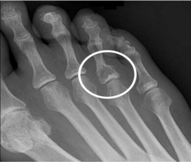
Similar to Non-ossifying Fibroma, Fibrous Cortical defect is usually asymptomatic. Many children and adolescents often do not even know that they have this defect present in their bones. This defect is discovered or diagnosed in a child or youngster incidentally through an X-ray for any other purpose. But some people who have experienced this disease reported the following symptoms.
- In the affected area, there is a soreness, discomfort, and mild swelling.
- Pain, slight cramps, and dull aches near the affected area, even without doing any work or activity.
After knowing these symptoms, the clinical experts perform a medical workup to examine the child’s bones’ condition and perform these diagnostic tests to reveal this disease if it is present.
- X-rays help in producing clear and sharp images of bones.
- MRI, which is the full form of Magnetic Resonance Imaging, is a very safe and genuine process without radiation exposure. This technique uses a combo of large magnets, a computer, and radio frequencies and presents a detailed image of muscles, bones, soft tissues, organs, and other parts within the body.
- Computed Tomography scan, an abbreviation of CT scan, uses the combo of a computer and X-rays that helps examine the structure of bones and presents images of the body in the form of slices (cross-sectional image).
- EOS, which is an imaging technology, creates and presents 3-D structure from 2 flat images. These images are taken when the child is in a standing position or an upright position. This technique enables improved and better diagnosis.
Fibrous Cortical defect Causes
Well, there are not many causes of this disease, but one cause of this disease is that it occurs due to the omission of the nest of fibrous tissue to calcify. Also, around 40% of children and adolescents who are skeletally immature have this defect present in their bones.
Fibrous Cortical defect Treatment
Fibrous Cortical defect does not require any treatment as this defect recovers on its own when the bones of children stop growing. But, in some cases, treatment is necessary to stabilize the defective bone after a fracture. There are many treatments present for bone tumors and other bone defects. The surgical treatments that are used in rare cases are defined as follows.
- Intralesional Curettage
In this procedure, the lesion is removed completely by scraping the bone.
- Bone Grafting:
In this procedure, the missing bone is replaced with either an artificial graft material or with the cadaver bone.
If any child has already experienced a broken bone, then, in that case, doctors may use casting or, they may surgically place pins and metal rods to give support to the bone and strengthen it.


