The term “silver fork deformity” is derived from the injured wrist having the appearance of an overturned dinner fork.it is also called colles fracture.
The distal (or lower end) radius is the most common site of fracture in the upper extremity. This is primarily seen in middle-aged and older patients, but it is not uncommon in any age patient. Distal radius fractures usually occur with a fall on an outstretched hand. These fractures may also occur in people who sustain a severe force to the wrist, such as a fall from a height, or motor vehicle accident. Combinations of these fracture patterns are also seen in children, which may involve the growth plate. A variety of fracture patterns may occur, but usually the distal radius compresses and displaces backwards producing the common appearance of a “silver fork deformity.”
Osteoporosis weakens the bones and makes the distal radius fragile and very susceptible to fracture. The vast majority of this type of fracture is successfully treated with closed reduction and casting. More recently, many unreducible or poorly reduced fractures have been treated with surgical reduction and internal fixation.
Colles fractures often heal just fine with proper splinting, usually by immobilizing with a cast. In some extreme cases, surgery might be necessary.
What is Silver Fork?
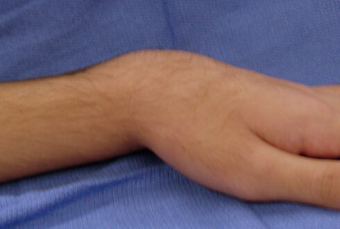
It is the deformity seen in Colles’ fracture in which the wrist or forearm has a curve like that of the back of a fork. Silver fork deformity is a wrist injury characterized by a distal radius fracture within 2.5 cm of its distal end. The arm will resemble a fork lying horizontally, so that the base of the fork’s tines makes an upward curve. The resulting hump in the fractured arm is due to dorsal displacement of the distal radius fragment.
Silver Fork Symptoms
In many cases a Colles’ fracture is an extraarticular, uncomplicated and stable fracture, but it can be intraarticular. Symptoms may include pain, swelling, deformity, and bruising. The symptoms may include the followings;
- Intraarticular radiocarpal or DRUJ extension of the fracture
- Radial shortening
- Loss of radial inclination
- Extension into the radiocarpal joint and the distal radioulnar joint
- Displacement of the articular fragments (blue arrow)
- Radial shortening and loss of radial inclination resulting in distal ulna abutting the lunate (yellow arrow)
- Fracture of ulnar styloid
- Pain with wrist motion or pressure on the wrist area
Silver Fork Causes
- Silver fork deformity usually results from a fall on an outstretched hand (FOOSH) which leads to a distal radius fracture.
- Usually caused by a broken radius bone (the big forearm bone on the same side as your thumb)
- have osteoporosis, a disease that weakens your bones
- are elderly
- have low muscle mass or poor muscle strength, or lack agility and have poor balance (these conditions make you more likely to fall)
- walk or do other activities in snow or on ice, or do activities that require a lot of forward momentum, such as in-line skating and skiing
- have an inadequate intake of calcium or vitamin D
Silver Fork Treatment
Silver fork deformity is treated with closed reduction it may be treated by following methods
- The most important treatment initially is immobilizing your wrist in a splint. You can simply use a magazine wrapped around your wrist to help support it. Elevate your wrist above the level of your heart to prevent further swelling. Putting an ice pack on the injury also helps reduce swelling.
- Over-the-counter medications such as acetaminophen and ibuprofen can help relieve pain.
- Don’t try to straighten your wrist, and avoid moving it around. Schedule an appointment with your doctor immediately, or go to an urgent care center for medical treatment. Go straight to the emergency room if the pain is severe or if your wrist is numb.
- Nonsurgical treatment; if your fracture isn’t serious, your doctor might place your wrist in a lightweight cast or splint and let it heal. They may need to straighten the bone if the fracture is displaced. This procedure, called a reduction, is done before your wrist is put in the cast. In most cases, the cast is taken off after a few weeks.
- Surgery; If your wrist is severely fractured, your doctor will recommend surgery to correct it. Your bones will be straightened and held together using pins, a plate and screws, or an external device that holds the pins in place. After surgery, you may need to wear a splint or cast to immobilize your wrist and help with pain relief.
- Physical therapy; Depending on the severity of your injury, you might have to work with a physical therapist or occupational therapist. You’ll do exercises to help rebuild strength in your wrist and regain your normal range of motion.


