Neuromas are clusters of nerve cells that are surrounded by Schwann cells in nearly equal numbers. Palisaded Encapsulated Neuroma is one of the most common neuromas characterized by the proliferation of nerve fibers without any prior injury. The other is the traumatic neuroma, also known as the amputated nerve, which is the proliferation of nerve fibers after trauma. The regenerative growth of nerve fibers is a feature of both of these kinds.
PEN (Palisaded encapsulated neuroma) is a malignant cutaneous tumor that appears as a mild, dome-shaped, flesh-colored lesion or papule. The lesions usually appear on the face, but they can also form on the limbs, trunk, or genital region, though this is uncommon. PEN is characterized by an intertwining network of benign spindle cells within the dermis.
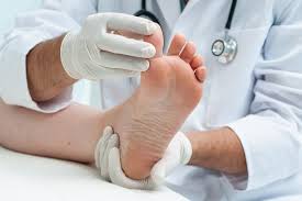
Palisaded Encapsulated Neuroma Symptoms
Oral lesions manifest clinically as a tiny ( 1 cm), moveable, submucosal, pink, asymptomatic nodule with slow growth.
Multiple lesions are uncommon in PENs. Patients often deny having experienced any past traumatic event. The lesion is characterized histologically by an overgrowth of Schwann cells with a diverse number and size of axons. The lesion is usually partially encapsulated by a thin capsule. The presence of S-100 protein elicits a favorable response from the cells.
PEN is distinguished by the presence of a papule that is 2–6 millimeters in diameter, sessile, firm, pink, or flesh-colored, and most often found on the face near mucocutaneous junctions. However, PEN can also be found in multiple numbers on the arms, oral mucosa, glans penis, eyelids, and acral sites.
Palisaded Encapsulated Neuroma Causes
The exact causes of Neuroma are unknown but there are certain contributing factors which are as follows:
- Neuromas develop as a result of biomechanical issues including a high or flat arch in one’s foot.
- This type of neuroma is mostly caused by certain trauma that affects the nerves.
- It is hazardous to wear shoes that are not properly fitted, as this might cause the toes to become compressed together. Avoid wearing shoes with heels that are more than two inches high. Shoes with this height can put too much pressure on the forefoot.
- A neuroma is caused or aggravated by repeated stress.
An atraumatic neuroma develops as a result of any injury to the nerve fibers in the peripheral nervous system. Injuries to the peripheral, somatic, or cranial nerves, as well as the autonomic nerves, frequently result in the development of traumatic neuromas. A traumatic neuroma is characterized by the development of a solitary cluster of regenerated axons and Schwann cells at the end of a nerve stump following nerve transection.
Palisaded Encapsulated Neuroma Diagnosis
PEN (Palisaded Encapsulated Neuroma) is diagnosed by a skin biopsy. When inspected under a microscope, pathological analysis of the biopsy reveals distinctive histological results. To aid in the diagnosis, the pathologist may employ specialized procedures such as immunohistochemical stains.
These neuromas resemble multiple endocrine neoplasia. Therefore, MEN 2B-associated cancers are evaluated clinically. Multiple clinical disorders share similar symptoms. To arrive at a conclusive diagnosis, the healthcare professional decides to conduct additional tests to rule out the possibility of other clinical diseases.
Palisaded Encapsulated Neuroma may resemble reactive soft tissue tumors clinically. Because it resembles other cancers with underlying systemic illness, its existence, especially at mucocutaneous junctions, needs a precise histologic diagnosis.
Palisaded Encapsulated Neuroma Treatment
Excision is recommended for traumatic neuroma, particularly for symptomatic pain relief. The nerve stump is repositioned into a scar-free region, such as inside a muscle flap. The use of 10 mg/cc intralesional kenalog before or post nerve repositioning surgery is employed.
To treat traumatic neuromas, surgical specializations such as orthopedic hand surgery or ENT surgery are included, depending on the tumor location. After a tissue diagnosis is established by the use of a biopsy, an MRI is frequently helpful in determining the degree to which other tissues are affected. Current surgical methods include removing a lot of nerves, fixing flaps, and then giving steroid injections.


