Uterine lipoleiomyoma is an extremely uncommon form of benign tumor. In 1991, Meis and Enzinger were the first people to describe the soft tissue tumor known as myolipoma. These tumors display typical histological characteristics, such as being made up of noncancerous smooth muscle and fully developed adipose tissue. Lipoleiomyomas are uterine tumors that resemble one another. Lipoleiomyomas mostly occur in the subserosal or any location of the uterus or cervix. Postmenopausal women are more likely to develop uterine lipoleiomyomas.
Lipoleiomyoma is an extremely rare uterine tumor that mostly affects obese perimenopausal and postmenopausal females. The tumor is made up of long bundles of smooth muscle cells that cross over each other and nests of fully grown fibrous tissue and fat cells.
Lipoleiomyoma is characterized by a fluctuating ratio of mature lipocytes to smooth muscle cells. In most cases, women who are asymptomatic, obese, and either perimenopausal or menopausal develop these tumors.
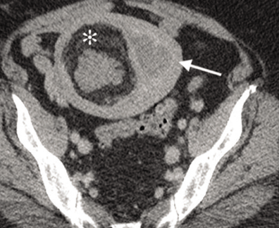
Imaging characteristics of lipoleiomyoma, which are offered by the tumor’s combination of fat, smooth muscle, and fibrous tissue, allow the radiologist to make a definitive diagnosis of the condition, despite its rarity.
Lipoleiomyoma Meaning
The lipoleiomyoma is a mass in the uterus that is not harmful. It is made up of fat cells, smooth muscle that is grown too much, and fibrous tissue. It is thought that this condition mostly occurs when uterine smooth muscle cells in a fibroid or leiomyoma change into fatty cells or die.
One particular type of leiomyoma is uterine lipoleiomyoma. A leiomyoma, which is also called fibroids, is a smooth muscle tumor that is usually harmless and rarely turns into cancer. According to histology, it is made up of smooth muscle cells and adipocytes in varying proportions, separated by thin fibrous tissue. The specific cause is unknown, but it is assumed to be fatty metaplasia of smooth muscle cells in a leiomyoma. Lesions range from a few millimeters and a few centimeters in size.
Lipoleiomyoma Symptoms
A palpable lump, heavy menstrual flow, and pelvic pain are common symptoms, much like they are with leiomyomas of the same size. Some people appear with symptoms like urine frequency, constipation, pelvic pain, and uterine bleeding, but many others show no signs at all.
Lipoleiomyoma of the uterus is recognized as a kind of leiomyoma. The symptoms of Lipoleiomyoma of the Uterine Corpus are typically determined by the tumor’s position, size, and whether it is a single or multiple tumour.
Some other symptoms are:
- Bleeding.
- A tumor’s internal bleeding causes tissue death.
- Hormone therapy, such as progesterone therapy, also causes bleeding or tumor tissue death.
- Discomfort during sexual activity.
- Abnormally high levels of circulating red blood cells as a result of the presence of a tumor.
- Hereditary leiomyomatosis and cancer of the kidney cells.
- Stomach fluid (ascites).
- Abdominal Enlargement.
- The abdomen gives the impression of being full if the patient possesses a large tumor.
- The constant need to urinate when the tumor presses on the urinary tract.
- Discomfort in the lower back.
- Abdominal pain and other signs and symptoms connected to the abdomen mostly occur in women who are expecting or using progesterone medication.
Lipoleiomyoma Causes
It is thought that uterine lipoleiomyomas are caused by the fatty breakdown or transformation of uterine smooth muscle cells inside a leiomyoma or fibroid. Leiomyomas of the ovary possess a mysterious origin in terms of their histogenesis. They likely come from smooth muscle cells in the blood vessels of the ovarian hilar, but they often also come from cells in the ovarian ligament, undifferentiated germ cells in the ovarian stroma, or smooth muscle cells. It is also believed that endometriotic cysts cause the surrounding stroma to transform into smooth muscle cells.
These tumors’ complex histogenesis, which often results from mesenchymal immature cells or the direct differentiation of smooth muscle cells into adipose tissues, is confirmed by immunocytochemical studies. Some lipid metabolic disorders and other situations related to estrogen insufficiencies, such as that which occurs during peri or post menopause, often induce abnormal intracellular fat accumulation.
Lipoleiomyoma Treatment
Lipoleiomyomas are clinically identical to leiomyomas and typically do not need treatment when they are tiny and asymptomatic. Therefore, it is essential to identify these tumours from ovarian teratoma, which is a condition that is only treated through surgical removal. Lipoleiomyomas are benign uterine tumours that do no impact mortality.
The treatment for these lesions is comparable to that for leiomyomas and is determined by the clinical symptoms as well as the size and location of the lesion. Embolization of the uterine artery or surgical excision is often performed, depending on the indication. In most cases, these tumours are completely harmless and possess positive outlook for treatment.


