Learn all about Epstein pearl symptoms, causes and treatment. Pearl Epstein, also known as gingival cysts, appears only in newborns. The study of American scientists from the National Institute of Health confirms the presence of Epstein pearls in 80 percent of newborns. Children who had just left the belly we must get used to the conditions in the new environment. Newborn skin is several times more sensitive than adults, no wonder, then, that appear on the various eruptions also in the mouth. Epstein pearls are whitish-yellow cysts. These form on the gums and roof of the mouth in a newborn baby.
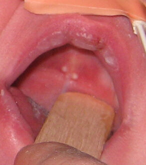
Epstein’s pearls were discovered by Alois Epstein in 1880. They are palatal cysts found along the median palatal raphe and arise from the epithelium entangled along the line of fusion. They are small white or yellow cystic vesicles (1 to 3 mm in size) often seen in the median palatal raphe of the mouth of newborn infants (occur in 60-85% of newborns). They are typically seen on the roof of the mouth (palate) and are filled with fluid. They are caused by entrapped epithelium (fissural cyst) during the development of the palate.
Epstein pearls disappear within 1 to 2 weeks of birth. These cysts arise from remnants of lamina. These are the nodules seen along median palatine raphe. Milia are a similar kind of skin problem in babies. Epstein pearls occur only in newborns and are very common. They are seen in about 4 out of 5 newborns. The condition was first described looking for online definition of Epstein pearls in the medical dictionary.
Epstein pearls are those white, pearly-appearing bumps (technically, cysts) found on the roof of the mouth or on the gums of about 80% of newborns. They are also technically known as gingival cysts of the newborn, but nobody uses that terminology.
They are distinctive, pearly white cysts that contain trapped mucous membrane type skin cells that slough off inside a closed pocket. The pearls found on the midline of the palate are undoubtedly formed when the halves of the palate fuse during early fetal development, trapping folded pockets of skin.
They cause no symptoms; they are often confused with candida infection in the mouth (thrush). They are generally shed within a few weeks probably as a result of friction with the nipple while nursing.
What are Epstein Pearls?
Epstein pearls are benign nodules, which range from one to three millimeters in size (less than a tenth of an inch) and appear on the roof of a baby’s mouth just behind her gums, are perfectly harmless. Similar-looking cysts can appear in other areas of your little one’s mouth, in which case they are known as Bohn’s nodules (also benign). These cysts contain epithelial cells (which act as a barrier to keep out dirt and microbes in the environment) as well as mucous membranes. You’ll find the same combination in the layers of moist tissue that line your baby’s urogenital, digestive, and respiratory tracts. As painful as they may look, Epstein’s pearls are painless good news for your baby.
Epstein Pearls Pictures
Epstein Pearls Symptoms
Gingival cysts, also called Epstein pearls, are small white-yellow raised bumps that appear on your baby’s gums or the roof of his mouth. The protrusions may look like budding baby teeth to new parents. Gingival cysts of the newborn rarely come singly, but appear in small clusters. They sometimes look like emerging teeth.
Epstein Pearls Causes
Even in the first trimester of pregnancy, the unborn baby’s jaw connects to the palate. In the process mucosa is “trapped” between the two which results in the appearance of just such a white-yellow efflorescence. The creation of pearls Epstein is therefore completely independent from good intentions we have and do not pose any threat to the health of her and her child.
Epstein Pearls Treatment
Epstein pearl treatment of any kind is not done, except for parental counseling and reassurance. It is important to note that management of all oral inclusion cysts (dental lamina cysts, Epstein pearls and Bohn’s nodules) remains the same, as all these have a self-limiting nature and require no treatment. Treatment of the smaller lesions includes careful wait and watch/surgical intervention, although spontaneous regression is rare. Larger lesions should be surgically excised under local or general anaesthesia at the earliest as they may risk the infant by causing feeding/respiratory difficulties. Congenital epulis needs surgical management in the majority of cases, whereas dental lamina cyst is self-limiting.


