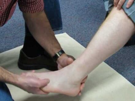
What is Peroneus tertius tendon?
The peroneus tertius tendon, also called the fibularis tertius tendon, is situated on the front of the lower leg. At the top, it is attached with the lower third of the fibula, one of the two bones of the lower leg, the peroneal ligament at the lower end connected to the metatarsal bone of the fifth toe. Sensory system supply are provided to this muscle by the fibular nerve. This muscle is missing in about 10% of the population. The arterial supply to the peroneus tertius tendon is an anterior tibial artery. PT capacity is to push the toes toward the shin (dorsiflexion) and move the foot away from the body’s center plane (eversion). Also, the PT muscle counters the rearranging power of the tibialis anterior, successfully leveling the foot.
Peroneus tertius tendon Tear
There are two peroneal ligaments; the peroneus longus and the peroneus brevis, which run corresponding to one another. Tendons are interfacing the outside of the foot to the peroneus longus and brevis muscles in your lower leg. These ligaments demonstrate how feet move outwards. They additionally help with balancing out your lower leg during weight-bearing exercises like running. Similarly, as with all instances of “tendonitis,” the issue is genuinely one of degeneration and injury. So a more appropriate term would be “peroneal tendinopathy” or “peroneal ligament dysfunction.”
A tendon rupture is a partial or complete tear of the tendon. Tendons are intense groups of tissue that join muscles to the bones. An incision might be brought about by a physical issue or expanded tension on the ligament during sports or a fall. Danger might be higher if one has a weak tendon, the peroneus tertius tendon, a short portion longitudinal split tear next to the peroneus tertius tendon’s inclusion.
Peroneus tertius tendon Pain
Symptoms associated with pain are burning sensation and swelling of the left ankle. There are no controlled clinical preliminaries that spread out a recovery program to follow. But some researchers suggested the treatment ought to incorporate rest. Issues with this muscle can show as the lower leg and heel torment.
Non-steroidal anti-inflammatory drugs (NSAIDS, for example, Ibuprofen) give relief from discomfort and irritation. Some activities include eversion practice each day, stretching out your calf muscles, and single-leg balance exercises are the best way to reestablish the tendon’s appropriate working. Other possible treatments like icing to be useful with peroneal tendonitis injury, similarly as with any tendon injury.
Peroneus tertius tendon Injury
Peroneus Tertius tendon injuries are very uncommon, with no recent detailed cases in history. Thus, there is little data relating to clinical importance, indicative moves, and remedial alternatives for PT injury or tear patients. But runners are more at risk of damages. Peroneal tendonitis makes up just about 0.6% of every running injury. Peroneus Tertius tendon presents as a sharp or hurting sensation along the tendon’s length or outwardly of your foot. It can happen at the insertion point of the tendons. Along the external edge of your fifth metatarsal bone or then again further up along the outside of your lower leg.
Activities that will be excruciating will attempt to dorsiflex and evert your foot, particularly against resistance. There may be some stiffness and soreness if patients do “lower leg circles” or, in any event, when latently extending the tendon. There shouldn’t be a lot of pain while standing or when the injured zone pushed softly. If the outside of the foot is delicate to touch, and on the off chance that the patient has a ton of pain standing or even while bearing non-weight, a patient may instead have a fracture on the fifth metatarsal.
Peroneus tertius tendon MRI
Magnetic resonance imaging (MRI) might be helpful to detect such kinds of problems. An uncommon high-grade tear injury of the peroneus tertius ligament was recognized, which has not been recently detailed in the literature. MRI is more often used in the peroneus tertius ligament sheath with a short section longitudinal split tear promptly near its insertion.
 Health & Care Information


